IMAGE

Fig. 1

ID
ZDB-IMAGE-240412-8
Source
Figures for Fasano et al., 2024
Image
Figure Caption

Fig. 1

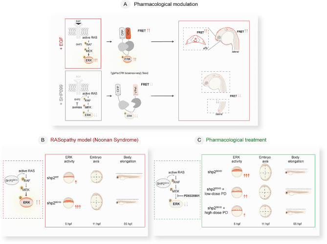
Schematics of the study design and outcome summarizing the main steps and outcomes of pharmacologically- and genetically induced RAS-MAPK pathway modulation assessed in Teen ERK reporter embryos. (A) Positive and negative modulation of RAS-MAPK signaling (via monitoring ERK activation) are obtained through pharmacological approach by acute stimulation with Epidermal growth factor, EGF, or prolonged exposure to the SPH2 inhibitor SHP099, respectively. ERK activation is assessed using the FRET-based ERK sensor Teen. In this study, an increase in the FRET signal in Teen embryos (due to elevated ERK activity) is visible in ventral forebrain upon EGF injection (upper panel). A decrease in FRET signal (due to reduced ERK activity) is visible in different brain domains and tail upon treatment with SHP099 (lower panel). (B, C) Genetic modulation of RAS-MAPK signaling in early embryos of a well-established Shp2D61G-NS zebrafish model (B) and partial rescue obtained by with low- (0.25 µM) and high-dose (1 µM) MEK inhibitor PD0325901 (PD) (C) are assessed by FRET in Teen embryos and precede onset of classical RASopathy morphological hallmarks (embryo axis and body length, validated at 11 hpf and 55 hpf, respectively). pERK: phosphorylated ERK

Acknowledgments
This image is the copyrighted work of the attributed author or publisher, and ZFIN has permission only to display this image to its users. Additional permissions should be obtained from the applicable author or publisher of the image. Full text @ Mol. Med.