
|
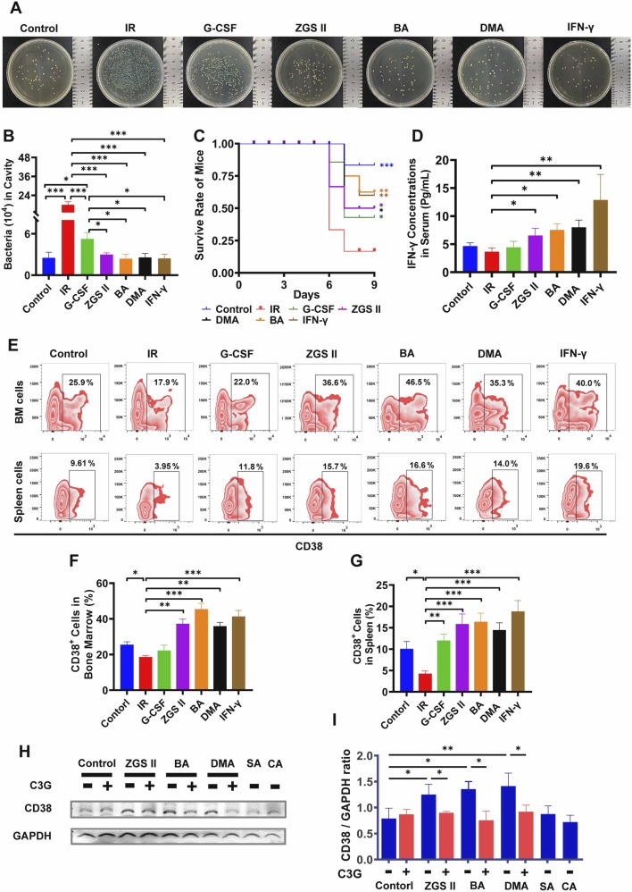
Fig. 7 ZGS II, DMA, BA, and IFN-γ enhanced bactericidal activity by inducing IFN-γ production in an irradiated mouse model. (A−B) Bacterial killing assay in the mouse cavity. The total colony number of extracellular viable bacteria in the cavity was counted at 16 h post-infection. (C) Leukopenia mice were intravenously injected with P. aeruginosa (6 × 10⁶ CFU per mouse) in the control group, model group, G-CSF-positive group (35 μg/kg), ZGSⅡ (10 mg/kg) group, BA (10 mg/kg) group, DMA (10 mg/kg) group, and IFN-γ (5 ×10⁴ U/mouse) group. Survival was observed for 4 days post-infection. Mortality differences compared with the infected model mice were statistically significant (* p < 0.05, ** p < 0.01, *** p < 0.001), as determined by a log-rank test. (D) Levels of IFN-γ serum were determined by immunoassay for each group on day 7. Data represent the mean ± SD of four mice in each group. * p < 0.05, * * p < 0.01, * ** p < 0.001 versus the model group (one-way analysis of variance). (E) Flow cytometry analysis of CD38 expression in BM cells and spleen cells for each group after treatment for 7 days. (F, G) The histogram represents the percentage of CD11b+ cells in BM (F) and spleen (G) for each group. (H, I) Western blot analysis (H) and quantification (I) of CD38 expression of NB4 cells treated with ZGSⅡ (10 μM), DMA (10 μM), BA (10 μM), SA (10 μM), CA (10 μM) or C3G (40 μM) for 5 days. Data represent the mean ± SD of three independent experiments. * p < 0.05, * * p < 0.01, * ** p < 0.001 versus the corresponding control groups (one-way analysis of variance); n.s.= no significance. BM: Bone marrow; IR, Irradiation.