
|
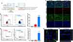
Fig. 4 Transcytosis efficacy of nanoformulations through the bEnd.3 cells and their uptake by SK-N-SH cells. (A) Schematic representation of the experimental design with the bEnd.3 cells monolayer cultured in the upper chamber (apical) of the insert Transwell® with 1 μm membrane pores and SK-N-SH cells seeded on the bottom of the basal compartment (the “brain side” of the Transwell®). Fluorescent formulations were added in the apical chamber at the equivalent concentration of 1.2 × 10¹⁰ particles and incubated for 24 h. Created with BioRender.com. (B) Flow cytometry plots indicating the percentage of nanoformulations (EVs-PKH26 and NPs-PLA-PEG-Cy5) taken-up by SK-N-SH after 24 h of incubation; the percentages of PKH26-positive or Cy5-positive cells are indicated inside the gates. (C) bar graph representing the quantification of the EVs-PKH26 (in red) and NPs-PLA-PEG-Cy5 (in blue) uptake and control cells (in black) were without nanoformulation. Data presented are mean ± SEM (n = 6). ***p < 0.001 vs control and ####p < 0.0001 vs control and EVs-PKH26. (D) Bivariate dot plots of PE (PKH26) and Cy5 fluorescence versus SSC of the nanoformulations (EVs-PKH26 and NPs-PLA-PEG-Cy5) present in the culture medium of the basal compartment of the Transwell®). (E) Quantification of nanoformulations in suspension in the basal medium after 24h of incubation. Data shown are mean ± SEM (n = 5), ****p < 0.0001 vs control and EVs-PKH26. (F) Representative fluorescence images showing the uptake of EVs-PKH26 and NPs-PLA-PEG-Cy5 by bEnd.3 cells (monolayer grown on the insert filter) after 24h incubation. EVs-PKH26 and NPs-PLA-PEG-Cy5 appear in red; core, blue; and claudin-5 (in green). Scale bar, 30 μm. Orthogonal views of 3D image stacks confirm the uptake of EVs-PKH26 (G) and NPs-PLA-PEG-Cy5 (H) in bEnd.3 cells. Scale bar, 60 μm.