IMAGE

Fig. 3.

ID
ZDB-IMAGE-240408-47
Source
Figures for Chen et al., 2024
Image
Figure Caption

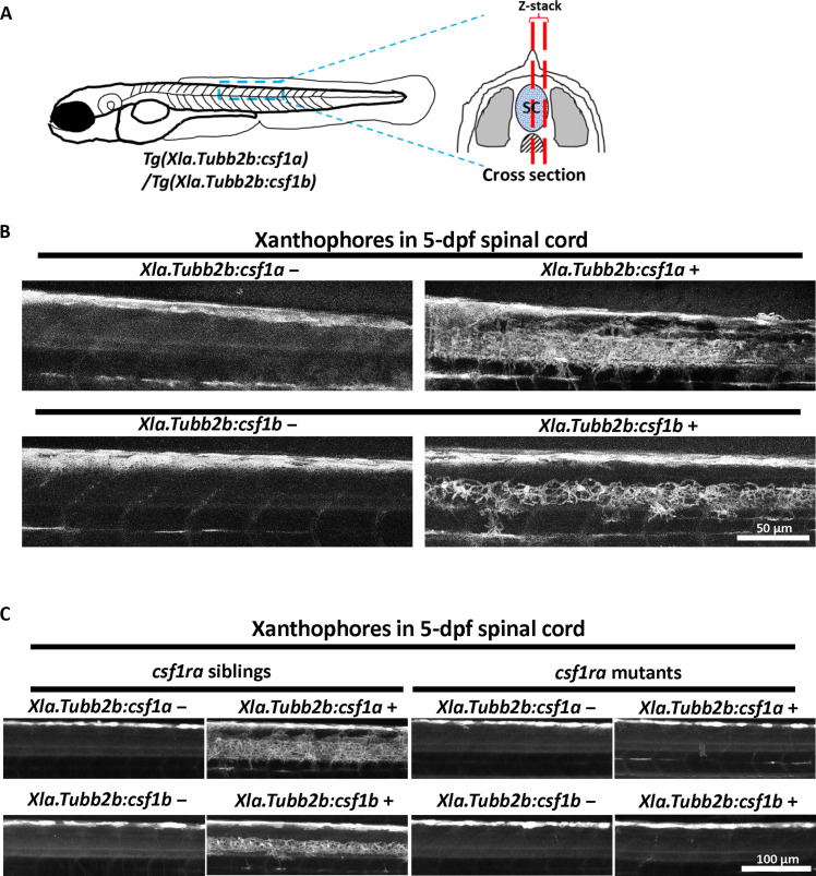
Fig. 3. Csf1a and Csf1b are chemoattractants of xanthophores.

(A) Schematic diagram of the imaging region. Blue dashed lines represent the imaging area at the dorsal trunk. Red dashed lines indicate the Z-stack range of confocal microscopy at spinal cord. SC, spinal cord. (B) Representative images of autofluorescent xanthophores at spinal cord region in 5-dpf larvae with (right) and without (left) Tg(Xla.Tubb2b:csf1a)/Tg(Xla.Tubb2b:csf1b). Xanthophores are found in the spinal cord region of transgenic larvae. (C) Transgenic and nontransgenic csf1ra mutants or siblings are imaged at 5 dpf to observe autofluorescent xanthophore. Images show spinal cord regions in respective groups.

Acknowledgments
This image is the copyrighted work of the attributed author or publisher, and ZFIN has permission only to display this image to its users. Additional permissions should be obtained from the applicable author or publisher of the image. Full text @ Sci Adv