
|
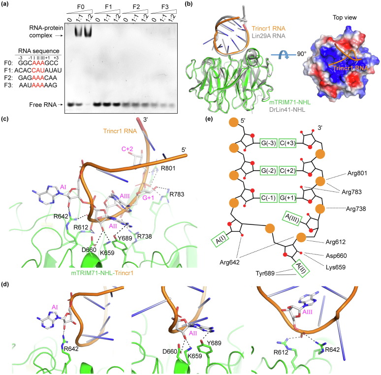
Fig. 1 Structure of mTRIM71-NHL complexed with Trincr1 RNA fragment. (a) EMSA results of mTRIM71-NHL bound to different RNA motifs of Trincr1 containing predicted hairpin structure (F0 and F1) or triple-adenosine bases (F2 and F3). Trincr1-F0 was shifted to a constant position by mTRIM71-NHL at low protein to RNA molar ratio, but Trincr1-F1, F2 and F3 was hardly shifted by mTRIM71-NHL. (b) Structure of mTRIM71-NHL bound to a 11 nt Trincr1 RNA (colored by green and orange), and its superposition to the structure of DrLin41-NHL-Lin29A (colored by gray). A top view of the electrostatic potential surface of mTRIM71-NHL and Trincr1 RNA was shown on the right panel. (c) Enlarged view of detailed interactions between mTRIM71-NHL and Trincr1 RNA. Sidechains of interacting residues and nucleotides are highlighted in a stick model, and hydrogen bonds are presented as dotted lines. (d) Detailed interactions of mTRIM71-NHL with AI, AII and AIII of the RNA individually. (e) Schematic diagram of interactions between mTRIM71-NHL and Trincr1 RNA. Hydrogen bonds are presented as dotted lines, and π-π stacking interaction is shown as solid line.