IMAGE

Figure 3

ID
ZDB-IMAGE-240315-45
Source
Figures for Liu et al., 2024
Image
Figure Caption

Figure 3

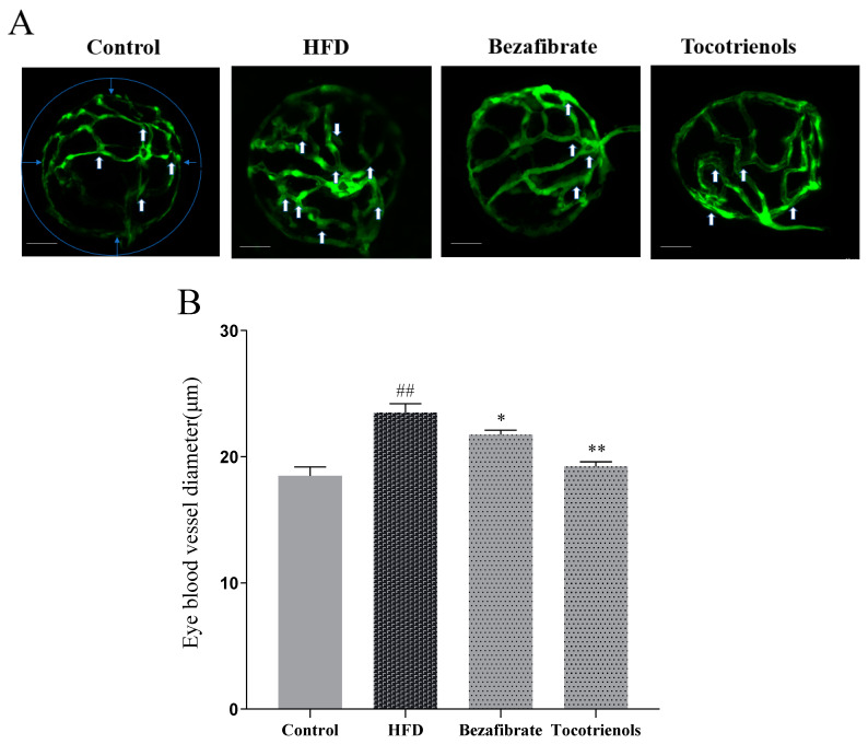
Changes in intraocular blood vessels in the zebrafish lens. (A) Confocal scanning of isolated Tg (fli1: EGFP) zebrafish eye, showing intraocular blood vessels in the lens. Transparent vessels can be seen from the outside through the translucent lens (indicated by white arrows in A), and the diameter of the intraocular blood vessels to be measured is indicated by blue arrows. Scale bar: 10 μm. (B) Quantification of the diameter of intraocular blood vessels in confocal scanning images, n = 6. ## indicates comparison with the blank control group, with p < 0.01. * indicates comparison with the model group, with p < 0.05. ** indicates a significant difference compared to the model group, with p < 0.01.

Acknowledgments
This image is the copyrighted work of the attributed author or publisher, and ZFIN has permission only to display this image to its users. Additional permissions should be obtained from the applicable author or publisher of the image. Full text @ Int. J. Mol. Sci.