IMAGE

Fig. 4

ID
ZDB-IMAGE-240315-40
Antibodies
Source
Figures for Töpf et al., 2024
Image
Figure Caption

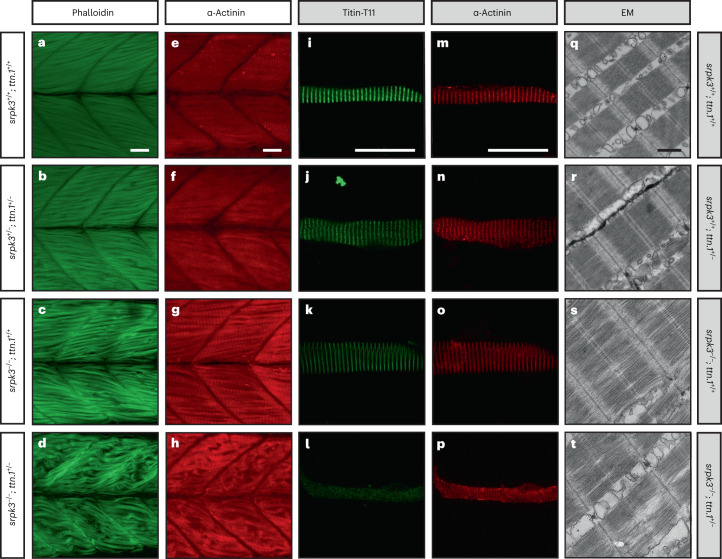
Fig. 4 ttn.1 heterozygosity induces a severe phenotype in homozygous srpk3-null mutant zebrafish larvae.

ah, Lateral view of Alexa Fluor phalloidin filamentous actin (green) and α-actinin Z-band marker (red) staining in skeletal fast muscle fibers in WT (a,e), srpk3+/; ttn.1+/ (b,f), srpk3/; ttn.1+/+ (c,g) and srpk3/; ttn.1+/ larvae (d,h) at 5 dpf. Compared to WT (a,e) or double heterozygotes (srpk3+/; ttn.1+/; b,f), homozygous srpk3-null alone only causes very mild muscle fiber defects (c,g), while ttn.1 heterozygosity in homozygous srpk3/ larvae severely affects muscle fiber integrity (d,h). it, Isolated myofiber immunostaining and electron microscopy (EM) in skeletal fast muscle fibers in WT (i,m,q), srpk3+/+; ttn.1+/ (j,n,r), srpk3/; ttn.1+/+ (k,o,s) and srpk3/; ttn.1+/ (l,p,t) larvae at 5 dpf. Isolated myofiber immunostaining showed that titin expression is largely reduced in the double mutant (srpk3−/−; ttn.1+/−; l,p) but not in the single heterozygous ttn.1 mutant (srpk3+/+; ttn.1+/−; j,n) or the srpk3-null (srpk3−/−; ttn.1+/+; k,o). EM showed that srpk3-null zebrafish (srpk3−/−; ttn.1+/+; s) had well-defined sarcomeres, with mildly disorganized myofibrils. The double-mutant fish (srpk3−/−; ttn.1+/−; t) displayed pronounced disruption of the sarcomere structure. White scale bars are 25 µm. Black scale bar is 500 nm. Representative images from >15 pooled fish per genotype.

Figure Data
Acknowledgments
This image is the copyrighted work of the attributed author or publisher, and ZFIN has permission only to display this image to its users. Additional permissions should be obtained from the applicable author or publisher of the image. Full text @ Nat. Genet.