
Image
|
Figure Caption
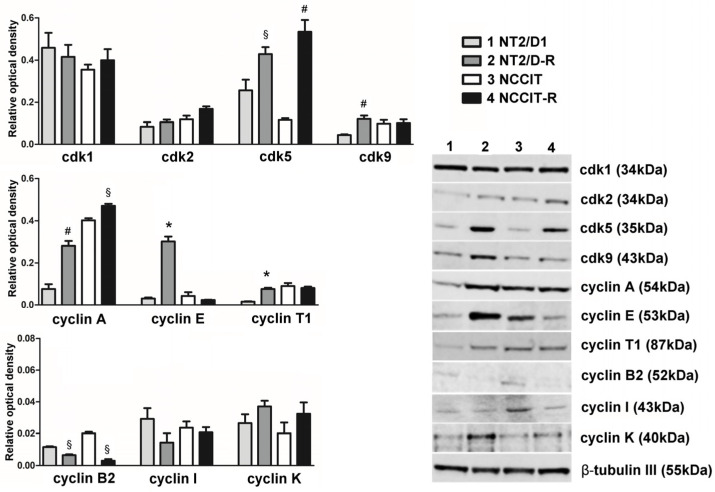
Figure 1
CDKs targeted by dinaciclib and their related cyclin protein expression in NT2/D1, NT2/D1-R, NCCIT and NCCIT-R cells. A total of 30 µg of total cell lysate was separated on a 4–12% Bis–Tris gel as described. Lane 1—NT2/D1, lane 2—NT2/D1-R, lane 3—NCCIT, lane 4—NCCIT-R. The human β-tubulin was used as an internal control. Quantification results are presented as a relative optical density means ± SEM of three independent experiments, and representative Western blot results are shown. *
Acknowledgments
This image is the copyrighted work of the attributed author or publisher, and
ZFIN has permission only to display this image to its users.
Additional permissions should be obtained from the applicable author or publisher of the image.
Full text @ Cells