IMAGE

Fig. 4

ID
ZDB-IMAGE-240229-147
Source
Figures for Lindahl et al., 2024
Image
Figure Caption

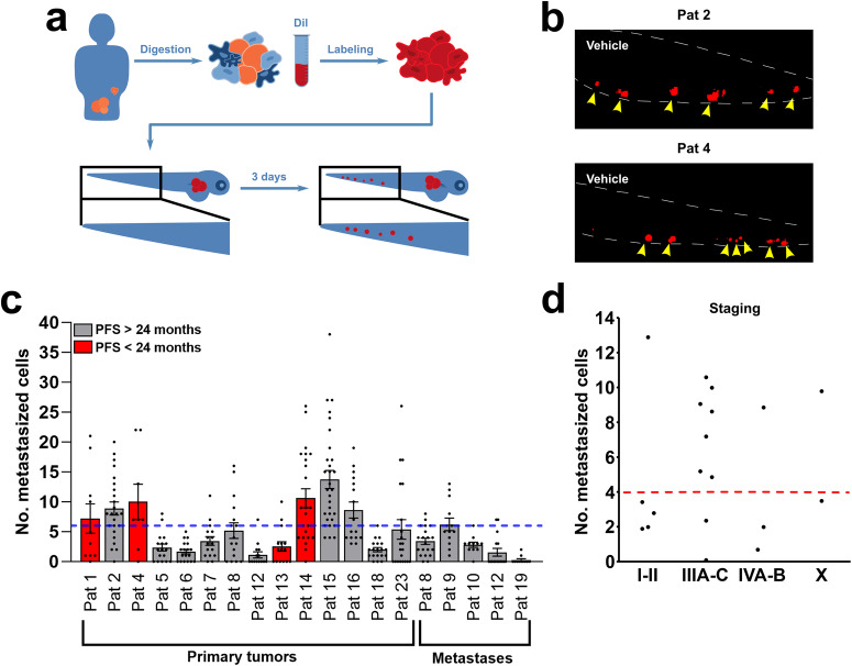
Fig. 4 Dissemination of tumour cells as a tool for prediction of outcome.

a Illustration of digestion, staining, and imaging of disseminated cells to caudal venous plexus (the area in the black box) of the zebrafish larvae. b Representative images of dissemination in the zebrafish larvae with tumour material from primary tumours of Patient 2 and Patient 4. Yellow arrowheads point to metastasized tumour cells (red). c Quantification of number of disseminated cells in the zebrafish larvae from primary tumours (n = 14, experiment done once) and metastatic lesions (n = 5, experiment done once) compared to patients who had a progression-free survival (PFS) of less than 24 months (red bars) with a cut-off value of 6 (blue dotted line). d Quantification of number of disseminated cells in the zebrafish larvae from primary tumours compared to staging with a cut-off value of 4 (red dotted line). n = 5, n = 9, n = 3, n = 2 for Stage I–II, Stage III A–C, Stage IV A–B, and Stage X (an initially unknown stage that was later confirmed to be Stage IV), respectively. All experiments were done once. Pat: patient.

Acknowledgments
This image is the copyrighted work of the attributed author or publisher, and ZFIN has permission only to display this image to its users. Additional permissions should be obtained from the applicable author or publisher of the image. Full text @ NPJ Precis Oncol