IMAGE

Fig. 4.

ID
ZDB-IMAGE-240229-122
Source
Figures for Mihalič et al., 2024
Image
Figure Caption

Fig. 4.

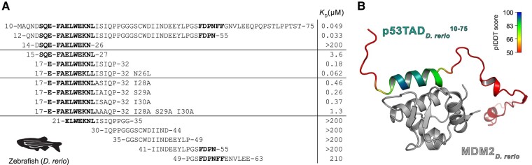
Affinities between p53TAD peptides and MDM2 from D. rerio. a) The overlapping peptides tiling a 51-residue stretch of p53TADD.rerio. To facilitate comparison, the numbering starts from the conserved motif and the residues of human p53TAD, F19xxxW23xxL26. With that in mind, the p53TAD10–75 in fact contains the whole TAD region of D. rerio and corresponds to p53TADfull-length. KD values for the peptide fragments were determined by the fluorescence polarization assay. Distal residues and Ile28Ser29Ile30 contribute to the affinity for MDM2. Both conserved motifs are in bold. The zebrafish silhouette was obtained from phylopic.org and is under CC0 1.0 DEED licence (https://creativecommons.org/publicdomain/zero/1.0/). b) ColabFold prediction of the interaction between D. rerio MDM2 and p53TAD10–75. The MDM2 is in gray, and the p53TAD10–75 is colored according to the pIDDT, where blue indicates highest and red lowest prediction confidence (pIDDT < 50).

Acknowledgments
This image is the copyrighted work of the attributed author or publisher, and ZFIN has permission only to display this image to its users. Additional permissions should be obtained from the applicable author or publisher of the image. Full text @ Mol Bio Evol