IMAGE

Fig 4

ID
ZDB-IMAGE-240226-112
Source
Figures for Baer et al., 2024
Image
Figure Caption

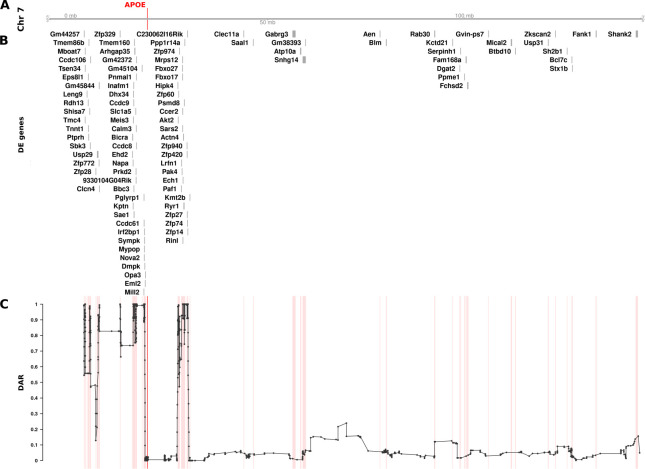
Fig 4 The relationship between DE genes and DAR along the entirety of mouse Chromosome 7 between male APOE2/2 and APOE3/3 mouse cerebral cortices (at 3 months of age).

The plot contains four sets of information represented by separate tracks horizontally. Track A represents the axis of Chromosome 7. The position of the APOE gene is marked and labelled in bold red. Track B displays differentially expressed genes according to their positions along the chromosome. 99 of 1126 total genes (8.79%) on Chromosome 7 that were expressed in the dataset were classified as DE (FDR < 0.05). Track C shows the trend in DAR as a connected scatterplot with each point in black representing the DAR value at a single nucleotide variant position (elastic sliding window size = 11 variants). Positions of the DE genes shown in track B are indicated by light red lines.

Acknowledgments
This image is the copyrighted work of the attributed author or publisher, and ZFIN has permission only to display this image to its users. Additional permissions should be obtained from the applicable author or publisher of the image. Full text @ PLoS Comput. Biol.