
Fig. 4
- ID
- ZDB-IMAGE-240223-34
- Publication
- Guillemyn et al., 2023 - Syntaxin 18 Defects in Human and Zebrafish Unravel Key Roles in Early Cartilage and Bone Development
- All Figures
- Figures for Guillemyn et al., 2023
|
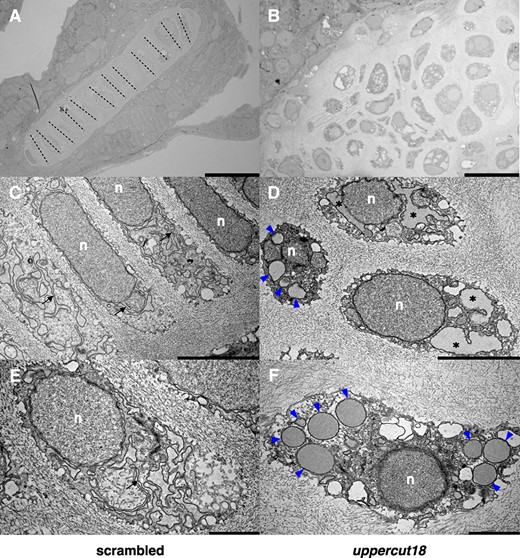
Fig. 4 Five‐dpf‐old uppercut18 zebrafish have compromised chondrocyte morphology and function. Electron micrographs of scrambled zebrafish (A, C, E) show cuboidal (polarized) chondrocytes that are in well‐organized stacks (dashed lines) and contain dense ER membranes that are well developed and organized (arrows), whereas uppercut18 zebrafish (B, D, F) are associated with irregularly formed and organized chondrocytes, distended ER (asterisks), and large amounts of electron‐dense material accumulating in the rough ER (blue arrowheads). n = nucleus. Scale bars (A–F) = 20, 5, and 2 μm, respectively. For A–F, 10 5‐dpf scrambled and crispant zebrafish were analyzed and representative samples are shown.