Image
Figure Caption

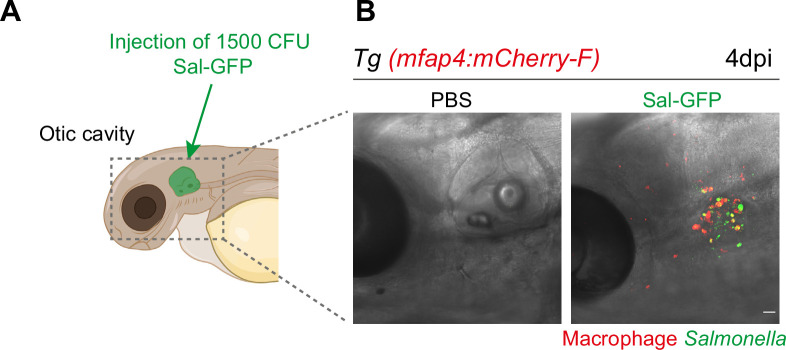
Figure 3—figure supplement 1. Macrophage response upon Salmonella injection in the otic vesicle.

(A) Schematic illustration of the otic vesicle injection site in 2 dpf zebrafish embryos. (B) Representative maximum projections of fluorescent confocal images of Tg(mfap4:mCherry-F) reporter embryos injected into the otic vesicle with either PBS- (left) or 1000 CFU of Sal-GFP (right), showing interactions of macrophages (red) and persistent Salmonella (green) at 4 dpi. Scale bar: 30 μm.

Acknowledgments
This image is the copyrighted work of the attributed author or publisher, and ZFIN has permission only to display this image to its users. Additional permissions should be obtained from the applicable author or publisher of the image. Full text @ Elife