IMAGE

Fig. 6

ID
ZDB-IMAGE-240202-7
Source
Figures for Larsson et al., 2024
Image
Figure Caption

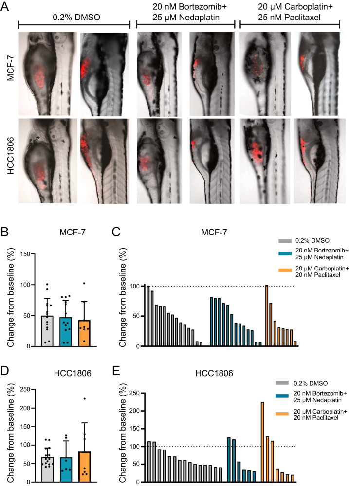
Fig. 6 Drug treatment of xenografted zebrafish larvae.

A Zebrafish larvae xenotransplanted with DiI-labeled (red) MCF-7 or HCC1806 human breast cancer cells were treated from 1–3 days post injection (dpi) with 0.20% DMSO, or a combination of 20 nM bortezomib and 25 µM nedaplatin or 20 µM carboplatin and 20 nM paclitaxel. Dot plots show relative change in tumor volume from baseline in MCF-7 treated with 0.20% DMSO (n = 14), 20 nM bortezomib and 25 µM nedaplatin (n = 13) or 20 µM carboplatin and 20 nM paclitaxel (n = 8) plotted as B mean with standard deviation C or individually. Dot plots show the relative change in tumor volume from baseline in HCC1806 treated with 0.20% DMSO (n = 15), 20 nM bortezomib and 25 µM nedaplatin (n = 6) or 20 µM carboplatin and 20 nM paclitaxel (n = 7) plotted as D mean with standard deviation E or as individual larvae. The dotted line indicates relative volume at 1 dpi.

Acknowledgments
This image is the copyrighted work of the attributed author or publisher, and ZFIN has permission only to display this image to its users. Additional permissions should be obtained from the applicable author or publisher of the image. Full text @ Cell Death Discov