IMAGE

Fig 3

ID
ZDB-IMAGE-240202-11
Source
Figures for Nielson et al., 2023
Image
Figure Caption

Fig 3

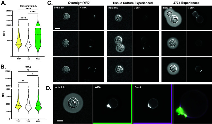
Yeast were incubated overnight in the indicated conditions, then isolated and fixed for microscopy at 63×/1.4NA. MECs have increased exposure to mannose and chitin despite the production of a thick capsule. Concanavalin (A) and wheat germ agglutinin (B) mean fluorescence intensities for Cn cultured in YPD, TC-experienced, or J774-experienced. Statistical comparisons represent the results of two-tailed, unpaired t-tests. (C) India ink and fluorescence images of YPD-cultured, TC-experienced, and J774-experienced Cn, three examples each. Staining in the first two conditions is punctate, at bud scars, or absent, while staining of J774-experienced cells is both punctate and circumferential. ConA is tagged with Alexa Fluor 633 (Fisher #C21402). Widefield images were taken with a 20×, 0.85NA objective. Exposures and intensity adjustments are uniform for all panels. (D) Example of ConA and WGA staining that overlaps on a different J774-experienced cell. Note intense staining at the bud scar with overlapping circumferential staining. WGA is tagged with Alexa Fluor 555 (Fisher #W32464). Widefield images were taken with a 20×, 0.85NA objective.

Acknowledgments
This image is the copyrighted work of the attributed author or publisher, and ZFIN has permission only to display this image to its users. Additional permissions should be obtained from the applicable author or publisher of the image. Full text @ mSphere