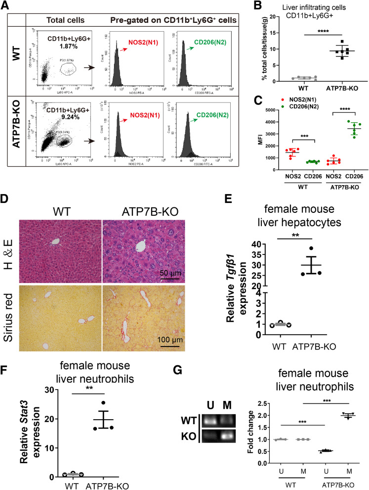
Fig. 19 The N2 neutrophils and liver injury phenotype also were observed in female ATP7B-KO mice. (A) Flow cytometric analysis of N1 (CD11b+Ly6G+NOS2+) and N2 (CD11b+Ly6G+CD206+) neutrophil populations in total CD11b+Ly6G+ cells derived from the livers of female wild-type and ATP7B-KO mice. (B) Flow cytometric quantification of CD11b+Ly6G+ neutrophils from female wild-type and ATP7B-KO mice livers. (C) Flow cytometric quantification of liver N1 and N2 neutrophils from female wild-type and ATP7B-KO mice. (B and C) n = 6 mice/group; unpaired 2-tailed t test. ∗∗∗P < .001, ∗∗∗∗P < .0001. (D) Pathologic changes in liver sections from female wild-type and ATP7B-KO mice at age 16 weeks. Upper panel: Representative H&E images in paraffin-embedded liver sections. Scale bar: 50 μm. Lower panel: Representative images of Sirius red staining in paraffin-embedded liver sections. Scale bar: 100 μm. (E) qPCR of mRNA levels of Tgfβ1 in liver hepatocytes isolated from female wild-type and ATP7B-KO mice. n = 3 sets of liver hepatocytes pooled from 2∼3 mice. ∗∗P < .01. (F) qPCR of mRNA levels of Stat3 in liver neutrophils isolated from female wild-type and ATP7B-KO mice. n = 3 sets of liver neutrophils pooled from 2∼3 mice. ∗∗P < .01. (G) Methylation-specific PCR in Socs3 promoter in liver neutrophils from female wild-type and ATP7B-KO mice. Left panel: Representative images of methylation-specific PCR in Socs3 promoter. Right panel: Quantification of band levels by ImageJ. M, methylated; U, unmethylated. n = 3 sets of liver neutrophils pooled from 2∼3 mice. ∗∗∗P < .001. WT, wild-type.
Image
Figure Caption
Acknowledgments
This image is the copyrighted work of the attributed author or publisher, and
ZFIN has permission only to display this image to its users.
Additional permissions should be obtained from the applicable author or publisher of the image.
Full text @ Cell Mol Gastroenterol Hepatol

