
Fig. 14
- ID
- ZDB-IMAGE-240130-30
- Publication
- Mi et al., 2023 - Stimulation of liver fibrosis by N2 neutrophils in Wilson's disease
- All Figures
- Figures for Mi et al., 2023
|
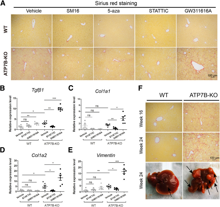
Fig. 14 The effect of N2 neutrophils on liver fibrosis in ATP7B-KO mice. (A) Representative images of Sirius red staining in paraffin-embedded liver sections from vehicle-, SM16-, 5-aza–, STATTIC-, or GW311616A-treated mice. Scale bar: 100 μm. qPCR of fibrogenic genes in (B) Tgfβ1, (C) Col1a1, (D) Col1a2, and (E) vimentin in wild-type and ATP7B-KO mice liver upon vehicle, STATTIC, or GW311616A treatment. n = 5∼6 mice/group. ∗P < .05, ∗∗P < .01, and ∗∗∗P < .001. (F) Upper panels: Representative images of Sirius red staining in paraffin-embedded liver sections from 16-week-old and 24-week-old wild-type and ATP7B-KO mice. Scale bar: 100 μm. Lower panel: Macroscopic liver pathology in 24-week-old wild-type and ATP7B-KO mice. WT, wild-type.