
|
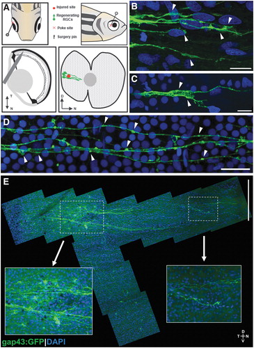
Fig. 1 Retina penetrating injury method and representative results. (A) Schematic of penetrating injury surgery procedure, and predicted injured site and regenerating RGCs with GFP expressed in gap43:GFP transgenic zebrafish. (B, C) Growth cones in regenerating RGCs marked by arrowheads. (D) Axonal varicosities (marked by arrowheads) in regenerating RGC axons. (E) Two-day postinjury retina flat-mount tiled image with magnified images of the injury site and the tip of regenerating RGC axons toward optic nerve head. Scale bars: (B, C) 10 μm; (D) 20 μm; (E) 200 μm. D, dorsal; GFP, green fluorescent protein; N, nasal; RGC, retinal ganglion cell; T, temporal; V, ventral.