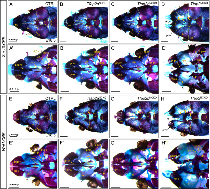
Formation of midfacial bone and cartilage structures depends on Tfap2a and Tfap2b gene dose in CNCCs. (A-H′) Alizarin Red (bone) and Alcian Blue (cartilage) staining preparations of E18.5 Sox10:CRE (A-D) or Wnt1:CRE (E-H) animals, with indicated genotypes and maxilla removed. n=5 per genotype. Scale bars: 1 mm. Anterior (a) is towards the left, posterior (p) towards the right. (A-H) Top-down views of the midfacial skeleton. Gold dashed lines (A-H) outline the peripheral edges of the calvaria bones. White dashed lines (B,F,G) outline nasal bone gaps. Gold arrowheads (D,F-H) indicate cartilaginous ectopias adjacent to the frontal bones. Asterisks (D,H) highlight missing nasal bones. (A′-H′) Bottom-up views of the midfacial skeleton. White arrowheads (B′,F′) indicate a gap in the vomer bones. cs, coronal suture; fr, frontal bone; na, nasal bone; nl, nasal/ethmoid labyrinth; ns, nasal septum; pl, palatine bone; pmx, premaxilla; ppp, palatal process of the palatine; pppm, palatal process of the premaxilla; ppro, pila preoptica; ppso, pila postoptica; ps, presphenoid; vm, vomer bone.
Acknowledgments
This image is the copyrighted work of the attributed author or publisher, and
ZFIN has permission only to display this image to its users.
Additional permissions should be obtained from the applicable author or publisher of the image.
Full text @ Development
Your Input Welcome
Thank you for submitting comments. Your input has been emailed to ZFIN curators who may contact you if
additional information is required.
Oops. Something went wrong. Please try again later.