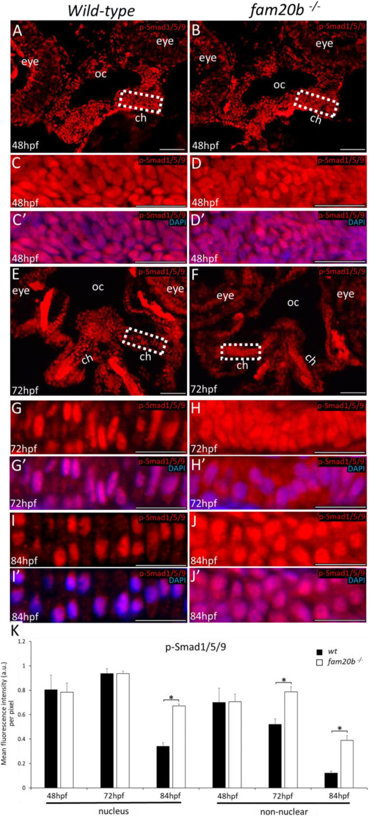
Smad-mediated BMP signalling is increased in fam20b−/− chondrocytes. (A-D′) Fluorescence immunostaining showed no clear differences between wild types (A,C,C′) and fam20b mutants (B,D,D′) in the levels of p-Smad1/5/9 in nuclei and non-nuclear regions of condensing ceratohyal mesenchyme at 48 hpf. (E-H′) p-Smad1/5/9 immunoreactivity appeared higher in non-nuclear regions of fam20b−/− ceratohyal chondrocytes (F,H,H′) at 72 hpf, compared with wild types (E,G,G′). (I-J′) p-Smad1/5/9 immunoreactivity appeared increased in both the nuclei and non-nuclear regions of fam20b−/− chondrocytes (J,J′), compared with wild types (I,I′), at 84 hpf. (K) Quantitative image analyses (n=3 for each group) demonstrated significant increases in p-Smad1/5/9 levels of fam20b−/− chondrocytes (*P<0.05, one-way ANOVA and paired Student's t-test). Scale bars: 50 μm. a.u., arbitrary units; ch, ceratohyal; oc, oral cavity; wt, wild type.
Acknowledgments
This image is the copyrighted work of the attributed author or publisher, and
ZFIN has permission only to display this image to its users.
Additional permissions should be obtained from the applicable author or publisher of the image.
Full text @ Development
Your Input Welcome
Thank you for submitting comments. Your input has been emailed to ZFIN curators who may contact you if
additional information is required.
Oops. Something went wrong. Please try again later.