IMAGE

Fig. 1

ID
ZDB-IMAGE-240122-12
Source
Figures for Guo et al., 2024
Image
Figure Caption

Fig. 1

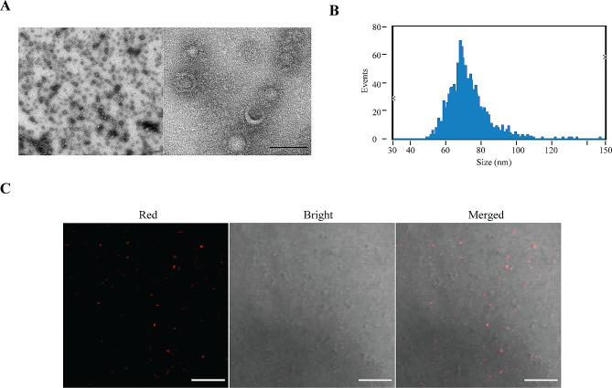
Characterization of P. gingivalis outer membrane vesicles (OMVs). (A) Transmission electron microscope (TEM) micrographs showing P. gingivalis OMVs. Electron microscopy imaging at 100 kv. Scale bar: 100 nm. (B) Size characteristics of P. gingivalis OMVs with a nanoparticle size analyzer (NSA). The dilution factor is 1000. The average diameter of P. gingivalis OMVs was about 72.28 nm with a concentration of 8.19 × 10¹⁰ particles/mL. (C) Confocal imaging results of P. gingivalis OMVs under 63x oil lens. Red, P. gingivalis OMVs labeled with PKH26. Scale bar: 10 μm

Acknowledgments
This image is the copyrighted work of the attributed author or publisher, and ZFIN has permission only to display this image to its users. Additional permissions should be obtained from the applicable author or publisher of the image. Full text @ BMC Oral Health