IMAGE

Fig. 3

ID
ZDB-IMAGE-231225-8
Source
Figures for Wang et al., 2023
Image
Figure Caption

Fig. 3

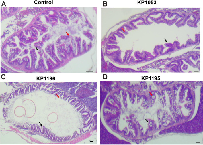
Changes in the structure of the gut 48 h after Klebsiella pneumoniae infected zebrafish with different virulence. The intestinal tract after infection was subjected to hematoxylin and eosin staining. A Control, (B) KP1052. C KP1196, and (D) KP1195. The red arrow indicates was goblet cells and the white arrows was epithelial cells. scale bars: 10μm

Figure Data
Acknowledgments
This image is the copyrighted work of the attributed author or publisher, and ZFIN has permission only to display this image to its users. Additional permissions should be obtained from the applicable author or publisher of the image. Full text @ BMC Infect Dis