Image
Figure Caption
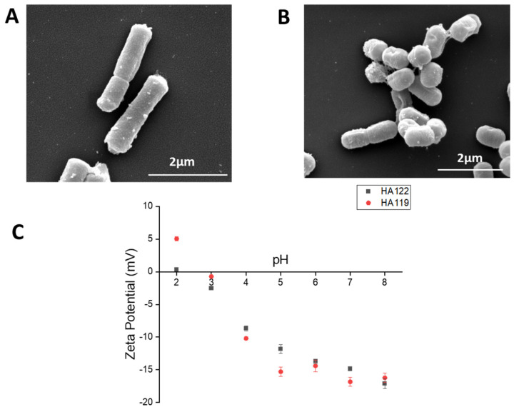
Figure 1
Morphology and surface charge of LHPost and LPPost. (
Acknowledgments
This image is the copyrighted work of the attributed author or publisher, and
ZFIN has permission only to display this image to its users.
Additional permissions should be obtained from the applicable author or publisher of the image.
Full text @ Microorganisms

