IMAGE

Figure 8

ID
ZDB-IMAGE-231225-121
Source
Figures for Zhai et al., 2023
Image
Figure Caption

Figure 8

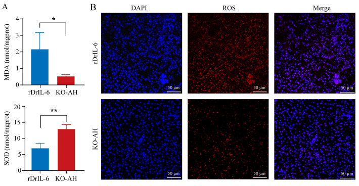
Effect of rDrIL-6 protein on liver oxidative stress in il-6−/− zebrafish post A. hydrophila infection. (A) MDA and SOD levels in liver tissues after A. hydrophila infection. Samples used for enzyme activity (biological replicates = 5, experimental repetitions = 3) were collected 12 h after infection. The experimental data were subjected to the Student’s t-test to identify the significance. Compared with the KO-AH group, the rDrIL-6 group showed significantly higher levels of MDA and lower SOD activities. * p < 0.05; ** p < 0.01. (B) Representative staining of ROS in liver sections. Frozen tissue sections were used for immunofluorescence detection of ROS. Red fluorescence corresponds to the presence of ROS, while blue fluorescence represents the cell nucleus. Scale bar = 50 μm. rDrIL-6: il-6−/− zebrafish treated with a mixture of A. hydrophila and rDrIL-6 protein; KO-AH: il-6−/− zebrafish treated with A. hydrophila.

Acknowledgments
This image is the copyrighted work of the attributed author or publisher, and ZFIN has permission only to display this image to its users. Additional permissions should be obtained from the applicable author or publisher of the image. Full text @ Int. J. Mol. Sci.