
|
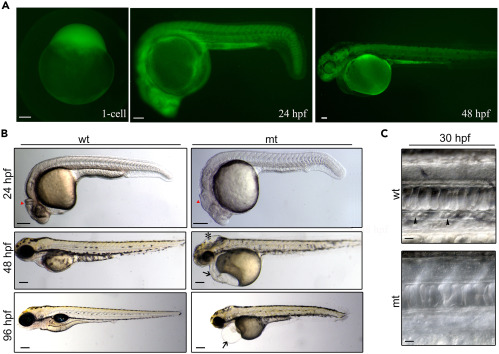
Fig. 1 GFP expression pattern and phenotypes of add1−/− transgenic embryos (A) Fluorescent images of add1−/− embryos. GFP was expressed maternally from the 1-cell stage and ubiquitously distributed along the whole body from 24 hpf to 48 hpf. Scale bars, 100 μm. (B) Morphology of add1−/− mutant and sibling embryos. At 24 hpf, MHB was poorly developed in the homozygous mutant embryos (red triangle). Swelling of the hindbrain was observed in homozygous mutant embryos at 48 hpf (black star). Black arrow showed the trimmer head and smaller eyes in homozygous mutant embryos at 48 hpf. The mutant embryos developed edema at 96 hpf compared to siblings. Scale bars, 100 μm. (C) Bright-field microscopy of blood cells in WT and mutant embryos at 30 hpf. Some blood cells could be observed in the caudal veins of siblings at 30 hpf (black arrowheads), whereas they were almost absent in the mutants. Scale bars, 10 μm.