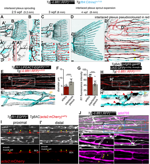
Fig. 6 Formation of the interlaced plexus at the fin base. (A-D″) Maximum intensity projections of confocal z-stacks of Tg(-0.8flt1:RFP)hu5333; Tg(flt4: citrine)hu7135 double-transgenic fish labelling all arterial ECs (black) and venous ECs (cyan). Scale bars: 30 μm for A; 50 μm for C; 100 μm for D. (A′,A″) Proximal fin plexus. Scale bar: 20 μm. (B,B′) Initial loops of the interlaced plexus at the fin base (pseudo-coloured in red in B′). Scale bar: 10 μm for B; 5 μm for B′. (C-D″) Interlaced plexus (pseudo-coloured in red). Blunt ends (C,C′, red arrowheads) and connections with bone enclosed arteries (D-D″, yellow arrowheads). Scale bar: 10 μm for C′; 20 μm for D′; 15 μm for D″. (E,E′) Maximum intensity projections of confocal z-stacks of Tg(fli1:LIFEACT-EGFP)mu240; Tg(-0.8flt1:RFP)hu5333 double-transgenic fish labelling all arterial ECs (red/cyan) and all ECs (white/black). Scale bars: 20 μm for E; 7 μm for E′. (F) Comparison of the diameters of bone-enclosed artery (BEA) and interlaced plexus (IP). Paired t-test (****P≤0.0001; n=17 segments from three individual fish for diameter measurements of the IP and BEA; data points indicate individual segments; data are mean±s.d.). (G) Flt1 fluorescence intensity measurements. Paired t-test (****P≤0.0001; n=30 cells for IP; n=32, data points indicate intensity values of individual cells; data are mean±s.d.). (H) Maximum intensity projections of confocal z-stacks of Tg(-0.8flt1:RFP)hu5333; TgBAC(pdgfrb:gal4ff;UAS:GFP)ncv24tg, nkuasgfp1a double-transgenic fish labelling all arterial ECs (red/cyan) and mural cells (white/black) in lateral views; anterior towards the left. (H′) Mural cells on the IP and BEA (red arrowheads). Scale bars: 10 μm for H; 5 μm for H′. (I) Maximum intensity projections of confocal z-stacks of Tg(Fli1:EGFP)y1; TgBAC(acta2:mcherry)ca8Tg labelling all ECs (white) and smooth muscle cells (red). (I) Proximal area. Scale bar: 50 μm. (I′) Distal area. Scale bar: 10 μm. (J-J″) Maximum intensity projections of confocal z-stacks of Tg(-0.8flt1:RFP)hu5333; qdot705 injections labelling arterial ECs (white) and blood vessel lumina (magenta). Yellow arrowheads indicate red blood cells. Scale bar: 20 μm for J; 7 μm for J′ and J″.
Image
Figure Caption
Acknowledgments
This image is the copyrighted work of the attributed author or publisher, and
ZFIN has permission only to display this image to its users.
Additional permissions should be obtained from the applicable author or publisher of the image.
Full text @ Development

