
|
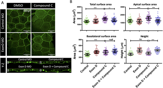
Fig. 5 Rab10-binding MyoVb isoform functions downstream of mTOR signaling during peridermal cell growth. (A) Confocal scans of lynEGFP-labeled peridermal cells of wild-type and Exon D morphant (Exon D MO) embryos treated with Compound C and DMSO at 48 hpf. (B) Graphical representation of the quantification of the total, apical and basolateral surface area, and the cell height in given genetic conditions and treatment. Data are median±interquartile range. **P<0.01, ***P<0.001 (Kruskal–Wallis test with Dunn's post hoc test). ns, not significant. ns#, although the difference is statistically not significant (P=0.097) the increasing trend might be biologically relevant (see the main text for details). Only key pairwise comparisons are shown in B; for the rest of the comparisons please refer to Table S4. Scale bars: 20 µm.