
|
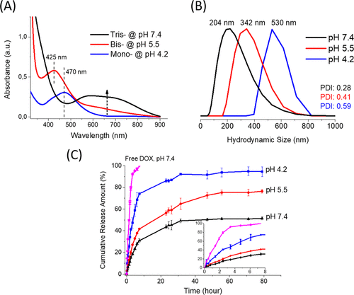
Fig. 6 (A) UV–vis absorption spectra of dopamine and V(III) mixture at different pH values: 7.4 (black), 5.5 (red), and 4.2 (blue) under argon gas. (B) DLS results of hydrodynamic size distributions of D-BSA NPs in PBS buffer at different pH values: 7.4 (black), 5.5 (red), and 4.2 (blue). PDI values are 0.28, 0.41, and 0.59 for pH (black), 5.5 (red), and 4.2 (blue), respectively. (C) Cumulative release amount of DOX from D-BSA NPs in 0.01 M PBS buffer at 37 °C at pH 7.4 (black), pH 5.5 (red), and pH 4.2 (blue). As a reference measurement, the release rate of the same amount of free DOX was also performed (pink). The initial 8 h of DOX release amounts from all samples were displayed in the inset of figure (C).