Image
Figure Caption
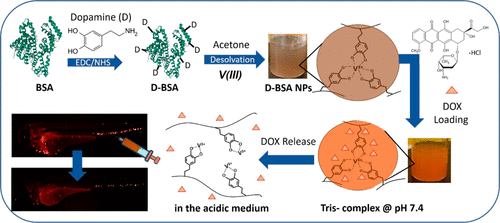
Fig. 1 Schematic representation of D-BSA and DOX-loaded D-BSA NPs formations. DOX was loaded on D-BSA NPs containing tris-catechol-V(III) coordination at pH 7.4, and DOX was released in the acidic medium upon degradation of NPs via formation of mono-catechol-V(III) coordination. Injection of DOX-loaded D-BSA NPs to zebrafish larval vasculature killed circulating tumor cells.
Acknowledgments
This image is the copyrighted work of the attributed author or publisher, and
ZFIN has permission only to display this image to its users.
Additional permissions should be obtained from the applicable author or publisher of the image.
Full text @ Biomacromolecules

