IMAGE

Fig. 6

ID
ZDB-IMAGE-231208-42
Source
Figures for Claude-Taupin et al., 2023
Image
Figure Caption

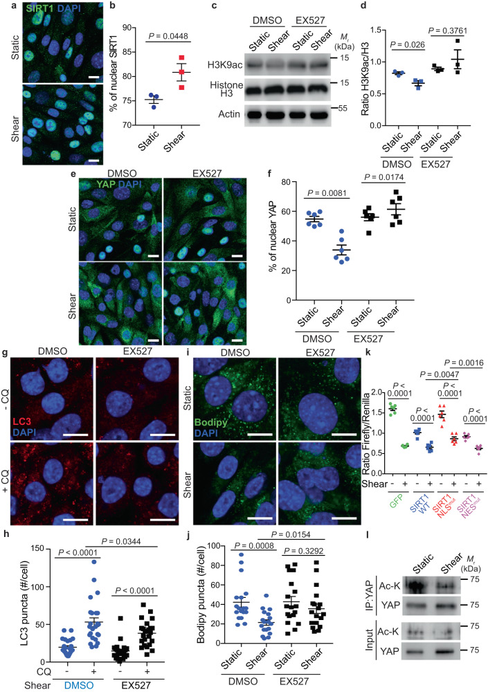
Fig. 6 SIRT1 induces YAP nuclear exclusion upon fluid flow.

a, b Representative images (a) and quantification (b) of SIRT1 nuclear localization in KECs subjected to shear stress during 24 h. Data show the mean ± s.e.m.; n = 3 independent experiments, two-sided t-test. Scale bars, 10 µm. c Representative images of H3K9ac, Histone H3 and Actin proteins levels in KECs subjected to flow (shear) or not (static) during 24 h, treated or not with EX527, by western blot analysis. d The ratio of H3K9ac to Histone H3 was determined by densitometry. Data show the mean ± s.e.m.; n = 3 independent experiments, two-sided t-test. e, f Representative images (e) and quantification (f) of YAP nuclear localization in KECs treated or not with EX527, subjected to flow (shear) or not (static) during 24 h. Data show the mean ± s.e.m.; n = 6 individual data points from 3 independent experiments, two-sided t-test. Scale bars, 10 µm. g, h Representative images (g) and quantification (h) of LC3 puncta in KECs subjected to flow (shear) or not (static) during 24 h, in the presence or absence of EX527 and chloroquine (CQ). Data show the mean ± s.e.m.; n = 24 individual data points from 3 independent experiments, two-sided t-test. Scale bars, 10 µm. i, j Representative images (i) and quantification (j) of lipid droplets (Bodipy) in KECs treated or not with EX527, subjected to flow (shear) or not (static) during 24 h. Data show the mean ± s.e.m.; n = 18 individual data points from 3 independent experiments, two-sided t-test. Scale bars, 10 µm. k Luciferase assay for YAP/TAZ activity in KECs transfected with GFP or GFP-SIRT1 WT or its mutated forms (SIRT1 NLSmut or SIRT1 NESmut), subjected to shear stress or not (static) during 24 h. Data show the mean ± s.e.m.; n = 6 from 3 independent experiments, two-sided t-test. l Immunoprecipitation analysis of YAP in KECs subjected to flow (shear) or not (static) during 24 h. Representative images of YAP and acetylated proteins are shown from n = 3 independent experiments. Source data are provided as a Source Data file.

Acknowledgments
This image is the copyrighted work of the attributed author or publisher, and ZFIN has permission only to display this image to its users. Additional permissions should be obtained from the applicable author or publisher of the image. Full text @ Nat. Commun.