
Image
|
Figure Caption
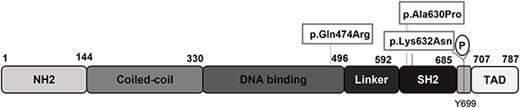
Fig. 1 Schematic of the human STAT5b protein adapted from Ramirez et al., 2020. The three variants studied are indicated: p.Gln474Arg located in the DNA binding domain, and p.Ala630Pro and p.Lys632Asn located in the SH2 domain. NH2, amino acids 1–144; Coiled-coil, amino acids 144–330; DNA binding, amino acids 330–496; linker, amino acids 496–592; SH2, amino acids 592–685; TAD, amino acids 707–787. Tyrosine 699 (Y699) can be phosphorylated by JAK2 and other kinases.
Acknowledgments
This image is the copyrighted work of the attributed author or publisher, and
ZFIN has permission only to display this image to its users.
Additional permissions should be obtained from the applicable author or publisher of the image.
Full text @ Hum. Mol. Genet.