
|
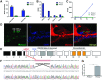
Fig. 4 Pknox2 expression in the inner ear and gene editing strategy. (A) Graph built using microarray data from manually collected OHCs and IHCs shows Pknox2 and Pknox1 expression in adult mice (Liu et al. 2014). (B) Graph built using RNA-Seq data for four types of cochlear cells showing Pknox2 and Pknox1 expression (Liu et al. 2018). (C) Pknox2 and Pknox1 expression is depicted from RNA-Seq data for GFP+ (hair cell–enriched) cochlear samples at four stages in developing mice (Scheffer et al. 2015). (D) Photomicrographs of immunofluorescence assays showing Pknox2 and Myosin 7 a expression in the inner ear of P8 wild-type mice. (E) Schematic of the PKNOX2 gene structure and the strategy developed to generate the mutant mice pedigree lacking Pknox2. The site of priming of the RNA guide on exon 4 and the STOP codon generated in exon 5 are indicated. Black boxes indicate 5′UTR exons. (F) Chromatograms of Pknox2+/+ and Pknox2−/− loci sequencing showing the deletion induced by Cas9 in the site of priming of the sgRNA guide. (G) Western blot quantification showing strong Pknox2 expression in wild-type mouse brain samples, a tissue where Pknox2 is strongly expressed. We observed the absence of expression in Pknox2−/− mice.