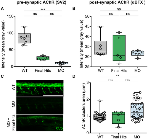
Fig. EV4 Additional AChR-related phenotypes in the gan zebrafish, which are not rescued by the five Final Hits A–D. Boxplots showing individual and mean values of the labeling intensity of presynaptic (stained with Synaptic Vesicle glycoprotein 2 (SV2)) (A) and postsynaptic (stained with α-bungarotoxin (αBTX)) (B) AChR clusters and area of post-synaptic AChR (D) for three groups: noninjected WT (black), MO-injected embryos (blue), MO-injected embryos treated with the five Final Hits (dark green), as identified in Fig 7. (C) Representative images of SV2 intensity labeling for noninjected WT embryos (WT), gan MO-injected embryos (MO) and gan MO-injected embryos treated with Phentolamine Hydrochloride. Analysis performed at 48 hpf. Scale bar represents a length of 500 μm. Each dot represents individual values for WT (n = 6 (A, B), n = 34 (D)) and MO (n = 6 (A, B), n = 43 (D)), and mean values of quadruplicate treated larvae (n = 5 (A, B, D)) with single Hits (A, B, D). The central bands of the boxplots represent the median, the boxes of the boxplots represent the interquartile range (between the first and third quartile), and the whiskers represent the minimum and maximum values. In the absence of normality of distribution of the data, a nonparametric Kruskal–Wallis test is applied; medians with range are represented; **P ≤ 0.01, ***P ≤ 0.001.
Image
Figure Caption
Acknowledgments
This image is the copyrighted work of the attributed author or publisher, and
ZFIN has permission only to display this image to its users.
Additional permissions should be obtained from the applicable author or publisher of the image.
Full text @ EMBO Mol. Med.

