IMAGE

Figure 1

ID
ZDB-IMAGE-231116-45
Source
Figures for Taler et al., 2023
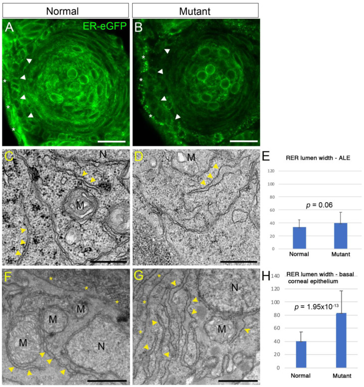
Image
Figure Caption

Figure 1

Abnormal rough endoplasmic reticulum (RER) in plod3 mutants. (A,B) Single confocal sections from eyes of 2 dpf normal (A) and mutant (B) embryos expressing ER-eGFP. Arrowheads point at the ALE. Asterisks mark the presumptive cornea. Scale bars are 20 µm. (C,D,F,G) Transmission electron microscope images of ALE cells (C,D) and basal corneal epithelial cells (F,G) from 2 dpf normal (C,F) and mutant (D,G) embryos. Arrowheads point at RER. Asterisks in (F,G) are located by the basal cell membrane, next to the developing corneal stroma in (F), which is missing in (G). M, mitochondrion; N, nucleus. Scale bars are 500 nm (C,D) or 1 µm (F,G). (E,H) Graphs depicting measurements of RER width from mutant and normal cells in the ALE (E) or basal corneal epithelial cells (H). Numbers on the Y axis are width in µm. Error bars are standard deviation. p values are depicted.

Figure Data
Acknowledgments
This image is the copyrighted work of the attributed author or publisher, and ZFIN has permission only to display this image to its users. Additional permissions should be obtained from the applicable author or publisher of the image. Full text @ Cells