IMAGE

Fig. 8

ID
ZDB-IMAGE-231113-33
Source
Figures for Park et al., 2023
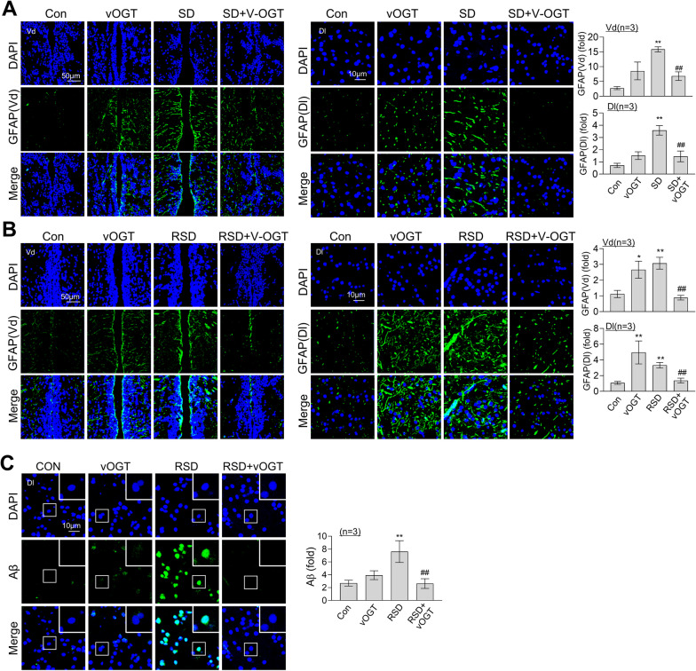
Image
Figure Caption

Fig. 8

Suppression of RSD-induced astrocyte activation and Aβ accumulation by overexpression of OGT. An vOGT virus was injected into the brains of zebrafish. Representative confocal images (40×) of DAPI (blue), GFAP (A and B, green), Aβ (C, green), and merged immunofluorescence staining in the Dl or Vd regions of zebrafish telencephalon are displayed. Enlarged images are presented in the white boxes. The graphs represent the quantitative results for each antibody with normalization by the DAPI levels (n = 3/group). For statistical analysis, the Kruskal–Wallis test with the original FDR of Benjamini and Hochberg was performed (*p < 0.05,  **p < 0.01 versus Con, ##p < 0.01 versus SD or RSD)

Acknowledgments
This image is the copyrighted work of the attributed author or publisher, and ZFIN has permission only to display this image to its users. Additional permissions should be obtained from the applicable author or publisher of the image. Full text @ J Neuroinflammation