
Image
|
Figure Caption
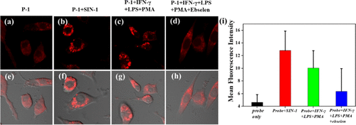
Fig. 6 Confocal fluorescence imaging of ONOO– in RAW 264.7 cells. (a) RAW 264.7 cells were incubated with P-1 (3 μM). (b) RAW 264.7 cells were loaded with SIN-1 (100 μM) for 30 min and then with P-1 for 1 h. (c) Cells were incubated with LPS (100 μM) for 4 h, IFN-γ (100 ng/mL) and PMA (10 nM) for 30 min, and then with P-1 for 1 h. (d) Cells were incubated with ebselen (100 μM) for 2 h, LPS for 4 h, IFN-γ and PMA for 30 min, and then with P-1 for 1 h. (e–h) Merged images. (i) Mean fluorescence intensities of panels (e–h). λex = 405 nm, λem = 550–620 nm.
Acknowledgments
This image is the copyrighted work of the attributed author or publisher, and
ZFIN has permission only to display this image to its users.
Additional permissions should be obtained from the applicable author or publisher of the image.
Full text @ ACS Biomater Sci Eng