
|
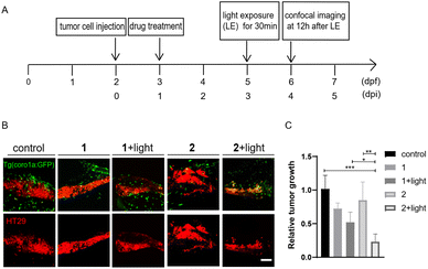
Fig. 8 Compound 2 displayed better antitumor effects than 1 in zebrafish xenografts. (A) The time scheme for the experiments in (B) and (C). 1 (10 μM) and 2 (10 μM) were added to the embryo medium accordingly. (B) Representative confocal images of HT29 tumor cells and innate immune cells from Tg (coro1a:GFP) zebrafish xenografts at 4 dpi treated with MSA-2, 1, 2 or 1 and 2 irradiated by light. (C) Relative tumor growth at 4 dpi versus 1 dpi. Results are shown as the mean ± SEM from multiple experiments, n = 6–9 embryos per group (***p < 0.001, **p < 0.01, *p < 0.05, t-test). Scale bar: 25 μm; dpf, days post fertilization; dpi, days post injection.