
|
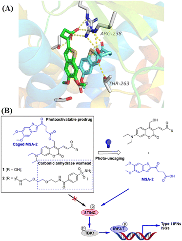
Fig. 1 Design of photo-activatable STING agonists. (A) Co-crystal structure analysis of MSA-2 bound to human STING protein (PDB: 6UKM26) identifies the terminal oxobutanoic acid as the critical structural motif for the activation of STING, evidenced by several key hydrogen bond interactions (orange dashed line). (B) STING agonist MSA-2 is caged with DEACM PPGs placed at the terminal oxobutanoic acid, which prevents the activation of the STING signalling. Irradiation with blue light at 450 nm removes the caging group to release MSA-2, which activates STING to induce the phosphorylation of the downstream signalling.