IMAGE

Figure 7

ID
ZDB-IMAGE-231031-14
Source
Figures for Li et al., 2023
Image
Figure Caption

Figure 7

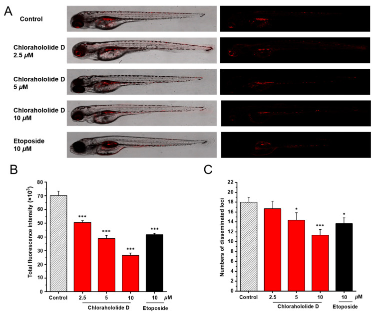
Chlorahololide D inhibited proliferation and migration in vivo. CM-DiI stained MCF-7 cells were microinjected into 2 dpf zebrafish embryos. After 4 h, tumor-bearing embryos were treated with chlorahololide D (2.5, 5, and 10 μM) for 48 h (n = 15/group). Etoposide (10 μM) was used as the positive control. (A) Intensity and distribution of the red fluorescence were photographed using a confocal microscope of disseminated foci in zebrafish. (B) The proliferation was quantified by ImageJ software. (C) The metastasis of MCF-7 cells was analyzed using ImageJ software. All of the results are expressed as the mean ± SD. * p < 0.05 and *** p < 0.001 vs. the control group.

Acknowledgments
This image is the copyrighted work of the attributed author or publisher, and ZFIN has permission only to display this image to its users. Additional permissions should be obtained from the applicable author or publisher of the image. Full text @ Molecules