IMAGE

FIGURE 2

ID
ZDB-IMAGE-231020-5
Source
Figures for Wei et al., 2023
Image
Figure Caption

FIGURE 2

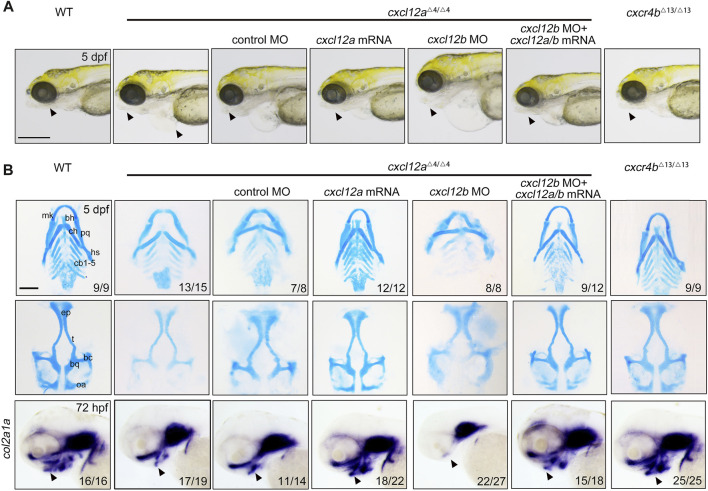
Depletion of cxcl12a impairs craniofacial cartilage development. (A) Phenotypic assessment of embryos at designated developmental stages, noting discernible craniofacial aberrations and pericardial edema in cxcl12a mutant embryos. The triangle annotates the zebrafish mandibular region. Scale bars set at 200 mm. (B) Alcian blue histochemical staining of craniofacial cartilage structures at 5 dpf and corresponding in situ hybridization images of col2a1a. Anatomical components denoted are: ep, ethmoid plate; tc, trabeculae cranii; mk, Meckel’s cartilage; bh, basihyal; ch, ceratohyal; pq, palatoquadrate; hs, hyosymplectic; cb, ceratobranchial. The triangle designates the mandibular sector. Scale bars: 100 µm.

Acknowledgments
This image is the copyrighted work of the attributed author or publisher, and ZFIN has permission only to display this image to its users. Additional permissions should be obtained from the applicable author or publisher of the image. Full text @ Front Cell Dev Biol