IMAGE

Fig. 1

ID
ZDB-IMAGE-231003-18
Source
Figures for Bradshaw et al., 2023
Image
Figure Caption

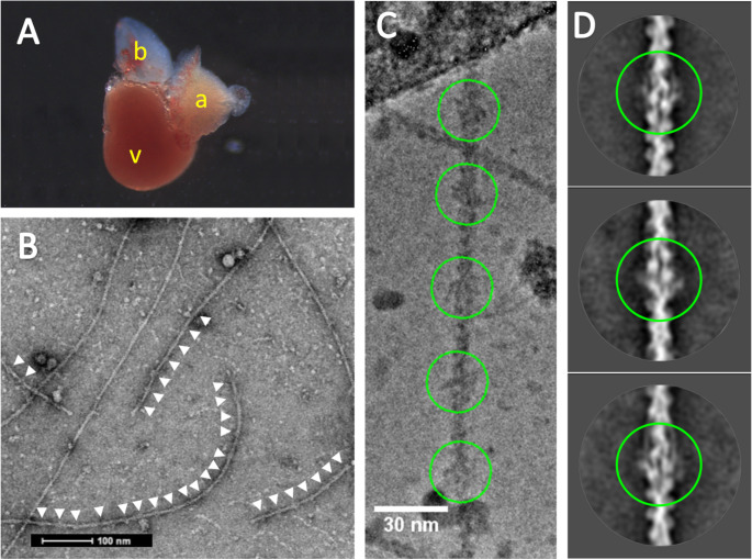
Fig. 1

Cryo-EM of zebrafish cardiac thin filaments. A: An excised adult Zebrafish heart, letters (in yellow) show V-Ventricle, A-Atrium B-bulbus arteriosus. B: A typical negative stain EM micrograph of isolated thin filaments, visible troponin complexes indicated with white arrows. C: Cryo-EM image illustrating the periodic binding of troponin on the thin filament (green circles) at a spacing of ~ 385Å D: 2D class averages from cryo-EM data with pairs of troponin labelling the two strands of actin (green circles); tropomyosin strands are also visible (top and bottom images)

Acknowledgments
This image is the copyrighted work of the attributed author or publisher, and ZFIN has permission only to display this image to its users. Additional permissions should be obtained from the applicable author or publisher of the image. Full text @ J. Muscle Res. Cell Motil.