
Fig. 2
- ID
- ZDB-IMAGE-231002-84
- Publication
- Tsai et al., 2023 - Endogenous tenocyte activation underlies the regenerative capacity of the adult zebrafish tendon
- All Figures
- Figures for Tsai et al., 2023
|
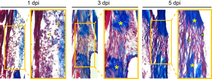
Fig. 2 Tendon regeneration proceeds through a rapid series of phases within the first week post-injury.
Masson’s trichrome staining of sections from regenerating tendons at 1 (left), 3 (middle), and 5 (right) dpi. Heavy infiltration of cells with myeloid-like morphologies can be seen at 1 dpi (yellow arrowheads). At 3 dpi, a fibroblastic bridge connecting the two severed tendon ends is evident. By as early as 5 dpi, the beginnings of collagen matrix deposition into the injury site are observed (green arrowheads). Yellow asterisks denote severed tendon ends and images were taken at 10x magnification. Dpi, days post-injury.