IMAGE

Figure 8

ID
ZDB-IMAGE-231002-199
Source
Figures for Fernandes et al., 2023
Image
Figure Caption

Figure 8

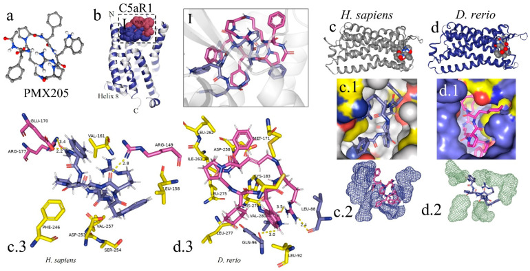
In silico analysis of the interaction of PMX205 with C5aR1 in D. rerio and H. sapiens. (a) 3D structure of PMX205 in the mol2 format of the ChemSpider database; (b) structural overlap of H. sapiens C5aR1 (grey) and homologous molecule of D. rerio (blue); (I) comparison of the PMX205–C5aR1 interaction between H. sapiens (purple) and D. rerio (pink); (c,c.1,d,d.1) docking of protein–binder interaction between C5aR1 and PMX205; (c.2,d.2) interaction of PMX205 with the binding site, in detail; (c.3,d.3) main amino acid residues of the receptors of H. sapiens (c.3) and D. rerio (d.3) and Van der Waals binding force with binder residues. The PDB C5aR1 file for D. rerio was built in the Swiss model, using the reference based on the crystallographic model 6C1R of H. sapiens, and the CB-Dock platform for docking.

Acknowledgments
This image is the copyrighted work of the attributed author or publisher, and ZFIN has permission only to display this image to its users. Additional permissions should be obtained from the applicable author or publisher of the image. Full text @ Int. J. Mol. Sci.