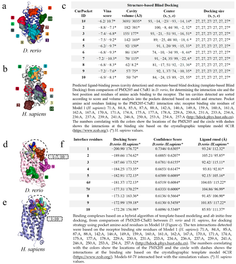
IMAGE

Figure 7

ID
ZDB-IMAGE-231002-197
Source
Figures for Fernandes et al., 2023
Image
Figure Caption

Figure 7

Virtual screening for the detection of the best D. rerio PMX205–C5aR1 interaction. (a) Docking showing specific interactions inside the drawn circle and non-specific ones outside the circle; (b) anchoring of PMX205-C5aR1 from H. sapiens showing model 1# specific interaction and other non-specific binding; (c) shows the top ten transients with regard to cavitation and blind docking; (*) H. sapiens values and (#) Model 1 selected through the position associated with PMX205 activity site based on crystallographic model 6C1R; (d) docking based on the amino acid sequence of Model 1# (Figure (c)), showing eight specific interactions inside the circle and two outside the circle representing nonspecific simulations from D. rerio; (e) for H. sapiens, all interactions were specific; (f) shows the values used for choosing the best interaction. (*) H. sapiens values. The numeric sequences refer to the colors that represent each docking interaction model.

Acknowledgments
This image is the copyrighted work of the attributed author or publisher, and ZFIN has permission only to display this image to its users. Additional permissions should be obtained from the applicable author or publisher of the image. Full text @ Int. J. Mol. Sci.