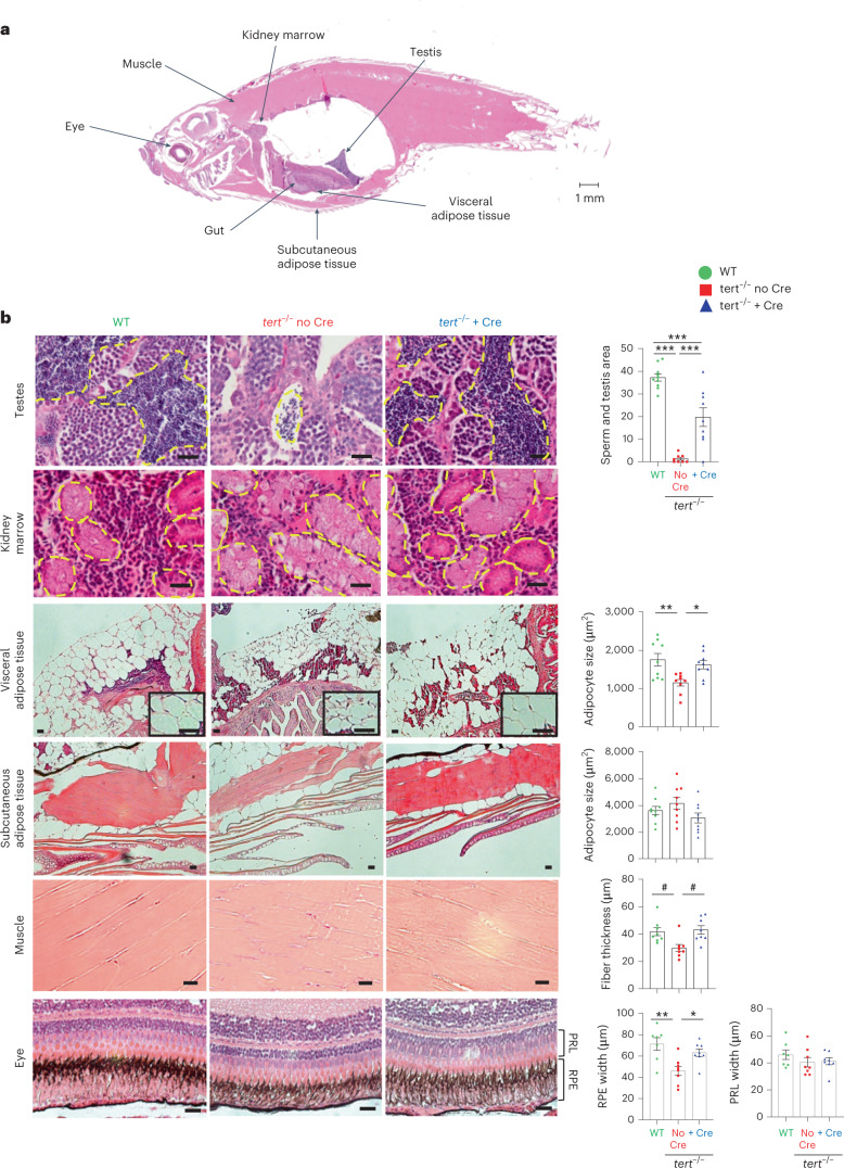
IMAGE

Fig. 4

ID
ZDB-IMAGE-230925-13
Source
Figures for El MaΓ― et al., 2023
Image
Figure Caption

Fig. 4

Gut-specific tert expression rescues systemic tissue degeneration.

Expression of telomerase in the gut of tert mutant fish rescued tissue degeneration in the testes, visceral adipose tissue, muscle and eye. a, Representative image of a longitudinal section of a zebrafish stained with H&E. The locations of each tissue analyzed in the study are indicated by arrows. b, Representative images of testes, kidney, visceral adipose tissue, subcutaneous adipose tissue, muscle and eye from 9-month-old fish stained with H&E (right). Except for the kidney, histological quantifications were performed for each tissue (left), namely the mature spermatids area (nWT = 10 fish, π‘›π‘‘π‘’π‘Ÿπ‘‘βˆ’/βˆ’noCre = 8 fish and π‘›π‘‘π‘’π‘Ÿπ‘‘βˆ’/βˆ’+Cre = 9 fish), adipocyte area (nWT = 9 fish, π‘›π‘‘π‘’π‘Ÿπ‘‘βˆ’/βˆ’noCre = 9 fish and π‘›π‘‘π‘’π‘Ÿπ‘‘βˆ’/βˆ’+Cre = 9 fish), muscle fiber thickness (n = 8 fish per group), retinal pigmented epithelium (RPE) and photoreceptor layer (PRL) (nWT = 7 fish, π‘›π‘‘π‘’π‘Ÿπ‘‘βˆ’/βˆ’noCre = 8 fish and π‘›π‘‘π‘’π‘Ÿπ‘‘βˆ’/βˆ’+Cre = 8 fish), respectively. Scale bar, 20 Β΅m. All data are presented as the mean Β± s.e.m.; *P < 0.05; **P < 0.01, ***P < 0.001, using a one-way ANOVA and post hoc Tukey test; *P < 0.05, using a Kruskal–Wallis test and post hoc Dunn test.

Source data

Acknowledgments
This image is the copyrighted work of the attributed author or publisher, and ZFIN has permission only to display this image to its users. Additional permissions should be obtained from the applicable author or publisher of the image. Full text @ Nat Aging