
|
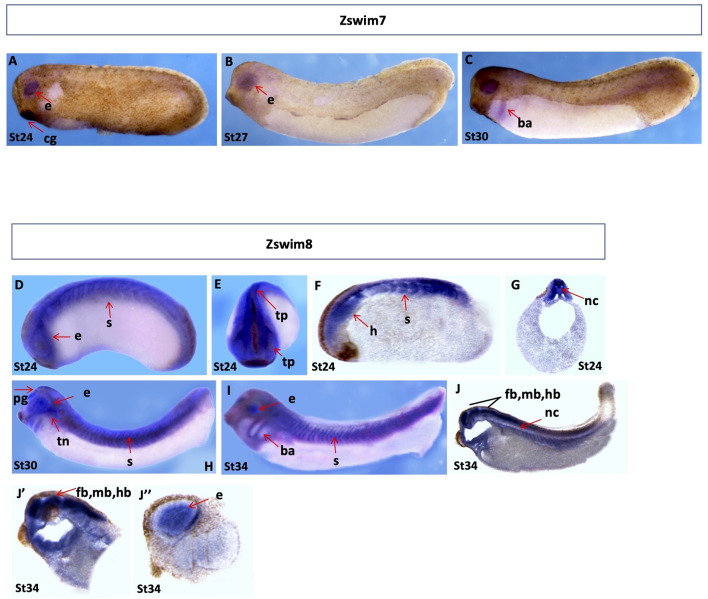
Fig. 7
Spatial gene expression patterns of zswim7 and zswim8
Spatial expression levels of zswim7 and zswim8 were analyzed by whole-mount in situ hybridization. Embryonic stages are indicated at the bottom of each image. A–C: Spatial expression of zswim7. Tailbud stages (stages 24 to 30), lateral view. D, E: Spatial expression of zswim8. D–G: Embryos at early tailbud stage (stage 24), lateral view (D), anterior view (E), longitudinal section (F), and transverse section (G). H, I: Embryos at tailbud stages (H, stage 30 and I, stage 34), lateral view. J–J’’: Longitudinal section of embryos at late tailbud (stage 34). b, brain; ba, branchial arch; e, eye; fb, fore brain; hb, hind brain; mb, mid brain; nc, notochord; pg, pineal gland; s, somites; tn, trigeminal nerves.