
Image
|
Figure Caption
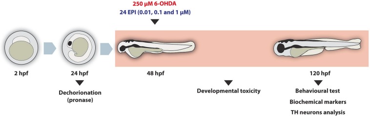
Fig. 1 Fig. 1. Exposure and analysis timeline. Fertilised eggs were collected at ∼2 h post-fertilization (hpf) and allowed to develop up to 24 hpf. At this point, the chorion was removed by pronase treatment and at 48 hpf animals were exposed for 72 h to 250 μM 6-OHDA, concomitantly to 24-EPI at different concentrations (0.01, 0.1 and 1 μM) and to 1 μM 24-EPI. Throughout the experimental period, developmental effects were assayed and, at the end of the exposure period, behavioural, oxidative-stress related parameters and TH-immunofluorescent neurons were evaluated.
Acknowledgments
This image is the copyrighted work of the attributed author or publisher, and
ZFIN has permission only to display this image to its users.
Additional permissions should be obtained from the applicable author or publisher of the image.
Full text @ Comp. Biochem. Physiol. C Toxicol. Pharmacol.