Image
Figure Caption

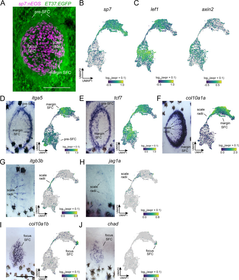
Figure 2—figure supplement 1. In situ hybridization using probes against specific transcripts localizes heterogeneous scale-forming cell (SFC) in the developing scale.

(A) Live imaging of differentiated SFC expressing sp7:nEOS (green) surrounded by a halo of pre-SFC labeled with ET37:EGFP (magenta). (B) UMAP visualization of sp7+ differentiated SFC and sp7- pre-SFC. (C) UMAP visualization of lef1 and axin2, transcripts localized to pre-SFC and margin SFC in previously published in situ hybridization (Aman et al., 2018). (D, E) itga5 and tcf7 were expressed in pre-SFC and SFC at the scale margin. (F) col10a1a was highly expressed in scale margin SFCs and radii SFCs but not pre-SFCs. (G, H) itgb3b and jag1a localized to radii SFCs. (I, J) col10a1b and chad were expressed predominantly in SFC at the scale focus. Scale bars, 50 μm.

Acknowledgments
This image is the copyrighted work of the attributed author or publisher, and ZFIN has permission only to display this image to its users. Additional permissions should be obtained from the applicable author or publisher of the image. Full text @ Elife