IMAGE

Fig. 6.

ID
ZDB-IMAGE-230916-281
Source
Figures for Lim et al., 2023
Image
Figure Caption

Fig. 6.

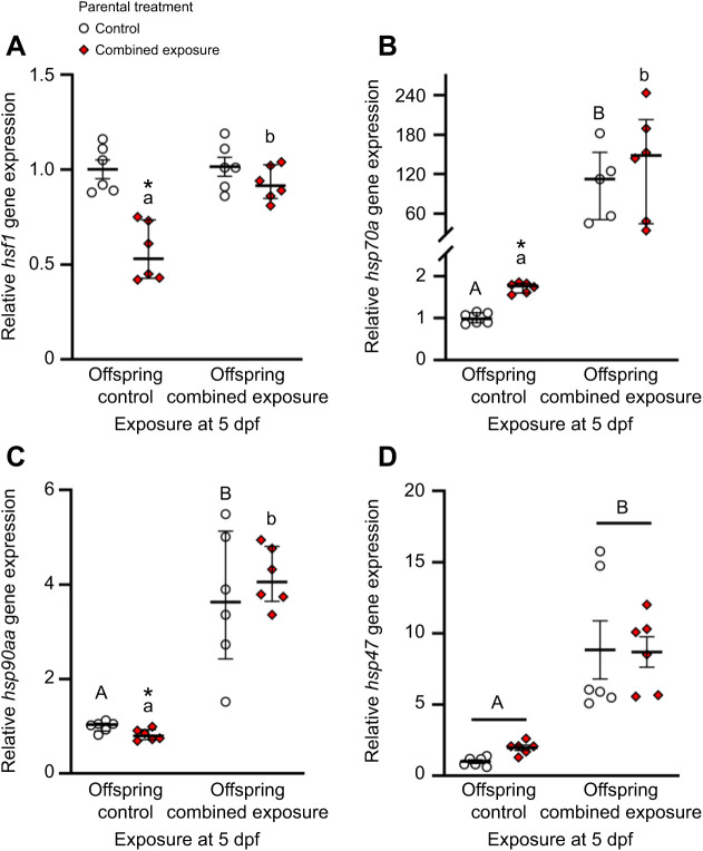
Effects of parental treatment on zebrafish larvae cellular stress gene expression. Control and post-exposure to combined elevated temperature and hypoxia relative gene expression for (A) hsf1, (B) hsp70a, (C) hsp90aa and (D) hsp47 in 5 days post-fertilization (dpf) larvae derived from adult zebrafish exposed to exposed to either control (offspring control) or combined exposure conditions (offspring combined exposure) for 14 days. Gene expression data were normalized and expressed as stated in Fig. 4. Statistical differences between values were determined by a two-way ANOVA followed by a Holm–Šidák post hoc test (D: square-root transformed, parental exposure: P=0.266; larval exposure: P<0.001; parental exposure×larval exposure: P=0.348), or two-tailed t-test (A: within control: P=0.830, within combined exposure: P<0.001) or Mann–Whitney rank sum tests (B: within control: P=0.004, within combined exposure: P=0.002; C: within control: P=0.002, within combined exposure: P=0.002) across exposure at 5 dpf, followed by two-tailed t-test (A: offspring combined exposure: P=0.174; B: offspring control: P<0.001, offspring combined exposure: P=0.484; C: offspring control: P=0.019, offspring combined exposure: P=0.477) or Mann–Whitney rank sum tests (A: offspring control: P=0.002) between parental treatments. Differences within treatments across exposure at 5 dpf are indicated by different letters. Differences between treatments for a given exposure at 5 dpf are indicated by an asterisk. Data are medians and interquartile ranges in A–C, and means±s.e.m. in D (all n=6; except hsp70a for combined exposure larvae from control parents, n=5).

Acknowledgments
This image is the copyrighted work of the attributed author or publisher, and ZFIN has permission only to display this image to its users. Additional permissions should be obtained from the applicable author or publisher of the image. Full text @ J. Exp. Biol.