IMAGE

Fig 6

ID
ZDB-IMAGE-230916-116
Source
Figures for Walker et al., 2023
Image
Figure Caption

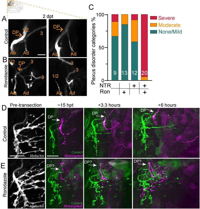
Fig 6 Schwann cells organize axon regeneration through the plexus.

(A) Maximum projection of the DP of zCrest:GFP to label abductor-specific axons at 2 dpt from 2 control animals. Schwann cells also express 37a> NTR-tagRFP. Nerves 1/2 and 3, the DP, and the abductor (Ab) and adductor (Ad) innervation are labeled. Both were categorized as “none/mild” plexus disorder. (B) Two examples of plexus disorganization category “severe” in animals with Schwann cells ablated after adding ronidazole. (C) Categorization of plexus organization in regenerated fins. Numbers indicate n. NTR = nitroreductase, Ron = ronidazole. (D, E) Timelapse imaging during regeneration of zCrest:GFP. (D) In the control, the plexus is organized with fascicles correctly targeting the abductor muscle (green) and incorrectly targeting the adductor muscle (magenta). Axons extend orderly projections into the fin musculature; n = 4/5 (E). When Schwann cells are ablated after adding ronidazole, axon growth through the plexus is disorganized and axons are defasciculated; n = 4/4. Scale bars are 25 microns (D, E) and 10 microns (A). Original data for panel C is in S1 Data. DP, dorsal plexus; dpt, day post transection; GFP, green fluorescent protein.

Acknowledgments
This image is the copyrighted work of the attributed author or publisher, and ZFIN has permission only to display this image to its users. Additional permissions should be obtained from the applicable author or publisher of the image. Full text @ PLoS Biol.